


Osteoarthritis (omarthrosis) of the shoulder joint is a chronic disease in which irreversible degenerative-dystrophic processes occur in the tissues of the joint. The pathology disrupts the normal functioning of the limb. The range of motion of the shoulder is gradually reduced to complete immobilityOsteoarthritis of the shoulder joint causes severe pain and reduces quality of life. Without treatment, disability can result.
In order to stop the processes of destruction of the joint and maintain mobility of the shoulder joint, it is necessary to contact an orthopedic specialist after the first symptoms appear.
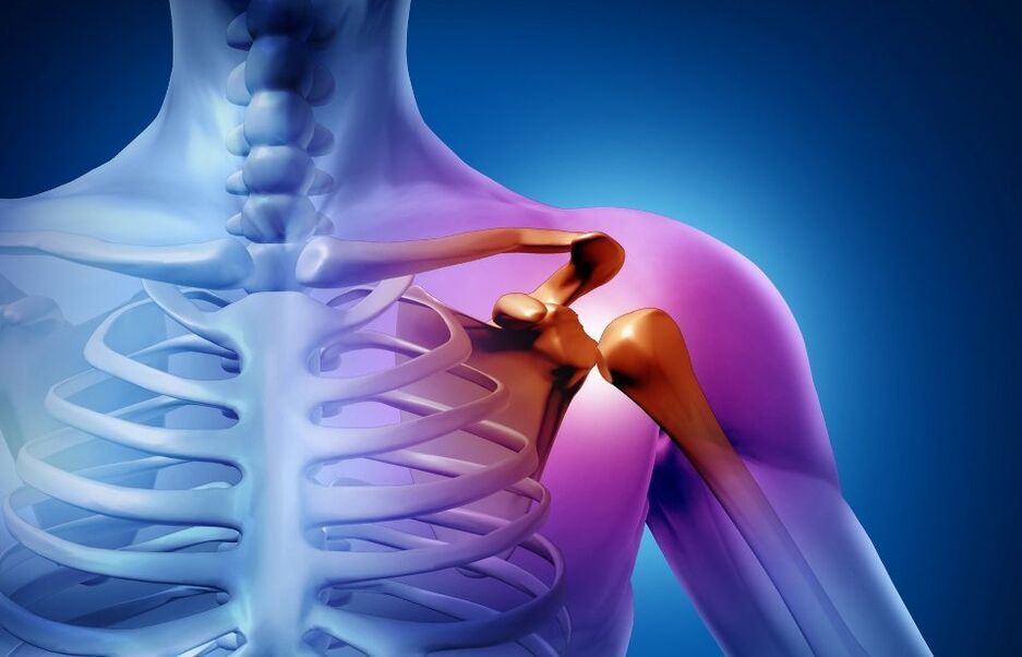
Osteoarthritis of the shoulder joint
The disease is polyetiological. The development of deforming arthrosis of the shoulder joint can be associated with various factors:
- Professional sports or intensive training.
- Endocrine diseases.
- Hormonal disorders.
- Congenital malformations of the development of the musculoskeletal system.
- hereditary predisposition, etc.
In most cases, secondary arthrosis is diagnosed: pathology occurs after exposure to the joint of one or another factor. Rarely register the primary, or idiopathic form of the disease. Tissue degeneration in this caseIt is impossible to establish the exact cause.
Symptoms of Shoulder Osteoarthritis
Changes in cartilage and bone tissue begin long before the first signs of arthrosis appear. Articular formations have a great potential for self-healing, therefore pathology is rarely diagnosed at a young age, when allMetabolic processes are significantly activated. As the body ages, recovery processes give way to degeneration. The first signs of destruction can appear after 40-50 years, and with the proliferative type of disease, patients have 16Changes are visible at the age of 18 years.
Shoulder Osteoarthritis Symptoms:
- Joint cracking during movement.
- Pain, especially severe after exercise.
- The rigidity of activity, expressed after sleep or long rest.
- The pain gets worse during the change of seasons.
degree of arthrosis
Clinical classification defines three degrees of arthrosis of the shoulder joint:
- 1 degree. The patient complains of a slight crunch that appears during movement. Pain syndrome is absent. Discomfort is felt when the arm is moved to the extremity position.
- 2 degrees. Pain occurs when the limb is raised above shoulder level. The range of motion is reduced. After excessive exertion, the patient feels pain even at rest.
- 3 degree. Joint mobility is severely limited. The pain syndrome is almost constant.
Diagnosis of osteoarthritis of the shoulder joint
The doctor needs not only to correctly diagnose, but also to determine the cause of the pathology. Treatment of the underlying disease significantly improves the patient's well-being and slows down cartilage degeneration.
manual examination
The first stage of diagnosis is a consultation with an orthodontist. The doctor examines the diseased joint for swelling, severe deformities. On the part of the development of arthrosis, the muscles may partially atrophy - this can be seen with the naked eyeIs.
With a manual examination, the doctor evaluates the function of the joint according to several criteria:
- The ability to voluntarily move hands.
- Thickening of the edges of the articular surfaces (larger osteophytes can be detected by palpation).
- The appearance of a crunch, a "click" that can be heard or felt with the arm during shoulder movement.
- Joint jamming in the presence of free chondromic bodies.
- Pathological movement in the shoulder.
radiograph
To detect signs of arthrosis of the shoulder joint, radiography is performed in two projections, which allow you to assess the degree of narrowing of the joint space, the condition of the bone surfaces, the size and number of osteophytes, the presence of fluidgives. and swelling of the surrounding tissues.
Ultrasound exam (ultrasound)
A non-invasive method that allows you to examine the joints in pregnant women and young children. According to the sonogram, the doctor determines the thickness of the cartilage, the condition of the synovial membrane. The method of osteophytes in the periarticular space, enlarged lymph nodesimagines well.
Magnetic resonance imaging (MRI)
The MRI machine continuously takes pictures of the sections. The images clearly show not only the joint, but also the adjacent tissues. To date, magnetic resonance imaging is one of the most informative methods in the diagnosis of arthrosis.
lab test
As part of a comprehensive examination, they appoint:
- General blood analysis. Based on the results, the doctor can judge the presence and severity of the inflammatory process. The analysis also helps to assess the general state of health.
- Analysis of urine. Kidney pathology often causes secondary deforming arthrosis. Analysis is essential for accurate diagnosis.
- Blood chemistry. The data helps to establish the cause of the inflammation. Biochemical analysis is also performed to monitor for complications and side effects during therapy.
Treatment of osteoarthritis of the shoulder joint
The therapy is long and difficult. The course of treatment includes medication, wellness procedures, a set of special exercises for arthrosis of the shoulder joint. In difficult cases, surgical intervention is indicated.
medical therapy
The drugs and dosage are selected individually. The doctor may prescribe:
- Non-steroidal anti-inflammatory drugs (NSAIDs). The drugs reduce inflammation and pain.
- Glucocorticosteroid preparations. Means based on hormones have a more rapid effect on the focus of pain. Drugs not only alleviate the patient's condition, but also reduce inflammation, exhibit antihistamine and immunosuppressive properties. Glucocorticosteroids in those casesare prescribed where NSAIDs are not effective.
- Painkillers. Drugs of this group are prescribed for severe pain syndrome. Depending on the severity of symptoms, the doctor selects non-narcotic or narcotic (rarely) analgesics.
- Chondroprotectors. The active ingredients of the drugs are involved in the formation of new cartilage tissue. The regeneration of the diseased joint is accelerated, trophism improves. Chondroprotectors have a cumulative effect and have proven themselves in the treatment of arthrosis of varying severity. has done.
Some drugs are injected directly into the joint cavity. For example, blockade has a better analgesic effect than taking the drug in the form of tablets.
physical treatment
Courses are carried out after removal of exacerbations. Physiotherapy as part of complex therapy helps to improve the transport of drugs in the diseased joint, relieve inflammation and reduce pain.
Use for the treatment of arthrosis:
- electrophoresis.
- Phonophoresis.
- Shock wave therapy.
Physiotherapy can be combined with massage, exercise therapy, therapeutic baths. It is best to undergo a set of procedures on the basis of a specialized clinic. The doctor will make a treatment plan taking into account the condition of a particular patient.
physical treatment
Moderate physical activity is important to slow down the degenerative processes. It is better to start exercise therapy for arthrosis of the shoulder joint in a medical center under the supervision of a doctor. The specialist will choose the exercises, teach them how to do them correctlyand the load is distributed so as not to cause aggravation of the disease. Gymnastics usually includes warm-ups, stretching and strength training. Exercises are performed at least 3 times a week.
After a course with a specialist, patients can perform therapeutic exercises for arthrosis of the shoulder joint at home.
Surgery
The operation is performed with arthrosis of the third degree, when the disease no longer allows the patient to move normally, causes severe pain, and prescribed therapy does not help.
There are several methods of surgical treatment:
- Perforation. A long needle is inserted into the joint cavity and the accumulated fluid is drained out. The puncture reduces pressure, reduces swelling, increases mobility of the joints. The procedure is minimally invasive, so it should be given an outIs carried out on an outpatient basis. The material obtained during the puncture is sent for research to determine the infectious agent or other indicators.
- Arthroscopy. With the help of microsurgery instruments, the doctor examines the joint cavity, removes scar tissue, makes a suture of the tendons of the rotator cuff or joint capsule if damaged. Many punctures remain on the skin. The patient recovers quicklygoes.
- Endoprosthetics. Endoprosthetics allows you to completely get rid of chronic pain, restore mobility of the hand. After the operation, long (from 3 to 6 months) rehabilitation is required.